Thalamus: The thalamus is a huge volume of gray matter within the dorsal part of the diencephalon of the brain, separated into two walnut-sized parts. Both of the thalami are found deep in the centre of the brain, between the midbrain and the cerebral cortex. The thalamus is a vital structure with several functions such as relaying of sensory signals, including motor signals, to the cerebral cortex, and the regulation of sleep, consciousness, and alertness. The thalamus may additionally be associated with the regulation of some types of memory. The thalamus not only transmits signals to the cortex but the cortex, in the rotation, sends signals back to the thalamus. 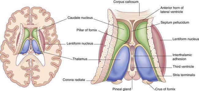 Injury to the thalamus can make coma. The most common injury to the thalamus produces central pain syndrome. This may be induced by hemorrhage when an artery through the brain is blocked.
Thalamus Anatomy
The thalamus a paired structure walnut-sized shaped of grey matter found in the forebrain that is superior to the midbrain, roughly the middle of the brain. The thalamus has four surfaces – Medial, lateral, superior and inferior surface and it has two ends or Poles -Anterior and posterior.
- Medial Surface: The medial surface of the thalamus forms the upper part of the lateral wall of the 3rd ventricle and is lined by ependyma. The medial surface is usually attached to the opposite thalamus by an interthalamic adhesion/bond. Inferiorly it is linked to the hypothalamus separated by an indistinct hypothalamic sulcus – this sulcus elongates from the upper end of cerebral aqueduct posteriorly to the interventricular foramen anteriorly. Through the junction of medial and superior surfaces, the ependyma of the third ventricle is revealed (from the lateral wall) to the roof. The line of reflection is indicated by a line designated taenia thalami, underlying which, there is a small bundle of fibers named stria medullaris thalami.
- Lateral Surface: The lateral surface of the thalamus is coated by a layer of myelinated fibers named external medullary lamina. The lamina divides lateral surface with the reticular nuclei.
- Superior surface: The superior surface of the thalamus is coated by a band of white matter designated stratum zonale.The medial part of this surface is separated from the overlying body of the fornix by the choroid fissure with the tela choroidea within it. More laterally it forms part of the floor of the lateral ventricle. The lateral border of the superior surface of the thalamus is signified by the stria terminalis and overlying thalamostriate vein, which divide the thalamus from the body of the caudate nucleus. Laterally, a slender sheet of white matter, the external medullary lamina, divides the main body of the thalamus from the reticular nucleus. Lateral to this, the thick posterior limb of the internal capsule occupies within the thalamus and the lentiform complex.
- Inferior Surface: The inferior surface of the thalamus is linked to the hypothalamus anteriorly and subthalamus posteriorly. The subthalamus divides thalamus from the midbrain tegmentum.
- Anterior end or Pole: It constitutes the posterior boundary of interventricular foramen.
- Posterior end or pole: Posterior end of the thalamus is designated as the Pulvinar. Pulvinar reaches beyond the third ventricle and overhangs the superior colliculus (Superior colliculus is a tiny elevation located on each side on the posterior aspect of the midbrain). Thus, pulvinar occupies just above and lateral to the superior colliculus. Subdivisions of Thalamus Inside, the thalamus is separated by a vertical Y-shaped of white matter, the internal medullary lamina within- Anterior nuclear groups Medial nuclear groups Lateral nuclear groups In extension, Intralaminar nuclei lie enclosed within and surrounded by, the internal medullary lamina. Reticular nuclei occupy laterally to the main nuclear mass, parted from it by the external medullary lamina. Midline nuclei either join the ependyma of the lateral walls of the third ventricle medially, or occupy adjacent to, and to some extent inside, the interthalamic adhesion. Medial and lateral geniculate bodies, previously regarded as Metathalamus, are now included as part of the Thalamus.
Connection of Thalamus The thalamus has multiple attachments to the hippocampus via the mammillothalamic tract, this tract holds the mammillary bodies and the fornix. The thalamus is linked to the cerebral cortex within the thalamocortical radiations.
The spinothalamic tract is a sensory pathway arising in the spinal cord. It sends information to the thalamus about temperature, pain, itch, and crude touch. There are two principal parts: the anterior (or ventral) spinothalamic tract, which carries crude touch and pressure and the lateral spinothalamic tract, which carries pain and temperature.
Thalamus Blood Supply
The thalamus receives its blood supply from plenty of arteries: paramedian thalamic-subthalamic arteries, the polar artery (posterior communicating artery), posterior (medial and lateral) choroidal arteries and inferolateral (thalamogeniculate) arteries. These are all derivatives of the posterior cerebral artery.
Thalamus Function
The thalamus is composed up of numerous collections of nerve cells that are centrally placed in the brain and are interconnected. Thalamus act as the relay station and integrate information into different subcortical areas of the brain and the cerebral cortex. There are thalamic nuclei concerning every sensory signal among the exception of the olfactory system. It takes sensory signals and sends them to the associated cortical area.
The medial geniculate nucleus of the thalamus transmits information connecting the inferior colliculus of the midbrain and the primary auditory cortex.
The lateral geniculate nucleus of the thalamus is associated with the visual system. It receives sensory inputs from the retina and acts rely upon station and process sensory information to the visual cortex. The medial geniculate nucleus acts as the relay station for the auditory system. The ventral posterior nucleus within the thalamus plays an important somatosensory relay that is liable for sending information relating to touch and proprioception to the primary somatosensory cortex.
Thalamus play a role in regulating sleep, wakefulness, and consciousness. The ventrolateral and the ventroanterior nuclei of the thalamus form part of the basal nuclei circuit and thus are committed in the performance of voluntary movements.
- Ventroanterior: It is concerned with the planning and initiation of movement.
- Ventrolateral: It is effective for the coordination and modulation of movement.
- Ventrointermedial: It supports coordinate different movements.
Thalamus Clinical Significance
The thalamic pain syndrome is caused by A cerebrovascular accident (stroke), which involves a one-sided burning or aching sensation often characterized by mood swings. Bilateral ischemia of the thalamus is supplied by the paramedian artery can produce serious difficulties including akinetic mutism, and be characterized by oculomotor problems. The occlusion of the Percheron artery can commence to a bilateral thalamus infarction.
Fatal familial insomnia is an inherited prion condition that is caused by the degeneration of thalamus, producing the patient to progressively lose his ability to sleep and advancing to a state of total insomnia, which habitually commences to death. In contrast, injury to the thalamus can occur in the coma.