Cerebellum: The cerebellum is a major feature of the hindbrain. The cerebellum performs an important part in motor control. It may additionally be associated while some cognitive functions such as attention and language as sound as in regulating fear and pleasure responses. The cerebellum resembles not initiate movement, but provides to precision, coordination, and accurate timing: it takes input from sensory systems of the spinal cord and from other components of the brain and integrates these inputs to fine-tune motor activity. Cerebellar damage produces disturbances in fine movement, equilibrium, posture, and motor learning in humans.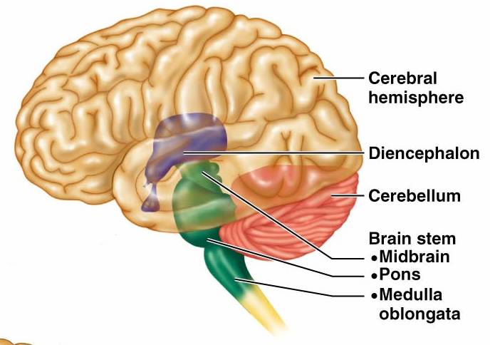
Anatomically, the cerebellum has the look of a separate structure attached to the bottom of the brain, tucked under the cerebral hemispheres.
Cerebellum Location
The cerebellum is positioned at the posterior part of the brain, directly under the occipital and temporal lobes, and inside the posterior cranial fossa. It is isolated from those lobes through the tentorium cerebelli, a tough layer of dura mater.
The cerebellum prevails at the alike level and posterior to the pons, from which it is divided by the fourth ventricle.
Cerebellum Anatomy
The cerebellum is divided into two hemispheres.which are connected by the vermis, a narrow midline area. Similar to the other structures of the central nervous system, the cerebellum consists of grey matter and white matter:
Grey matter found on the surface of the cerebellum. It is tightly folded, making the cerebellar cortex.
White matter found under the cerebellar cortex. Set in the white matter are the four cerebellar nuclei (emboliform, the dentate, globose, and fastigi nuclei).
The cerebellum comprises innumerable neurons than the total from the rest of the brain but takes up just 10% of the total brain volume. The number of neurons in the cerebellum is linked to the number of neurons in the neocortex. There are approximately 3.6 times as many neurons in the cerebellum as in the neocortex.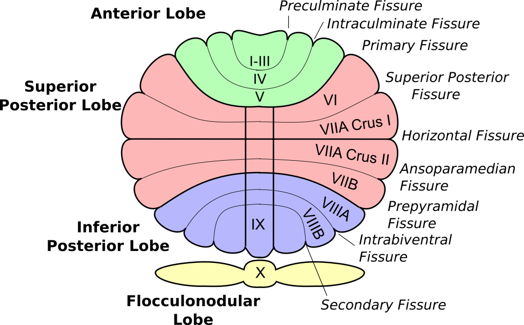
Anatomical Lobes of the cerebellum
There are three anatomical lobes in the cerebellum; the anterior lobe, the posterior lobe and the flocculonodular lobe. These lobes divide the cerebellum from rostral to caudal. These lobes are separated by the primary fissure and posterolateral fissure. The flocculonodular lobe is often called the vestibulocerebellum.
Zones of the cerebellum
There are three cerebellar zones. In the midline of the cerebellum is the vermis. Each side of the vermis is the intermediate zone. Lateral to the intermediate zone holds the lateral hemispheres. There is no variation in gross structure among the lateral hemispheres and the intermediate zones
Functional Divisions of the cerebellum
The three functional areas of the cerebellum – the cerebrocerebellum, the spinocerebellum and the vestibulocerebellum.
- Cerebrocerebellum – the largest division of the cerebellum, made by the lateral hemispheres. Cerebrocerebellum is involved in planning movements and motor learning. It takes inputs from the cerebral cortex and pontine nuclei and transmits outputs to the thalamus and red nucleus. The Cerebrocerebellum area also regulates coordination of muscle activation and is essential in visually guided movements.
- Vestibulocerebellum – the functional equivalent to the flocculonodular lobe. Vestibulocerebellum is concerned with controlling balance and ocular reflexes, principally fixation on a target. It gets inputs from the vestibular system and transmits outputs back to the vestibular nuclei.
- Spinocerebellum – including the vermis and intermediate zone of the cerebellar hemispheres. It is implicated in regulating body movements by supporting error correction. It additionally receives proprioceptive information.
Blood Supply of the cerebellum
The cerebellum takes blood supply from three paired arteries:
- Superior cerebellar artery (SCA)
- Anterior inferior cerebellar artery (AICA)
The SCA and AICA are branches of the basilar artery, - Posterior inferior cerebellar artery (PICA)
The PICA is a branch of the vertebral artery.
Venous drainage by the superior and inferior cerebellar veins. They drain toward the superior petrosal, transverse, and straight dural venous sinuses.
Cerebellum Function
The cerebellum gets information from the sensory systems, the spinal cord, and other components of the brain and later coordinates motor movements. While the cerebellum is not considered initiating movement, but it regulates such as posture, coordination, balance, and speech, resulting in a smooth and well-balanced muscular activity.
The cerebellum plays a significant role in this motor learning behavior.
Cerebellar Disorder
Damage to the cerebellum, while not producing paralysis or intellectual impairment, might commence to a lack of balance, tremors (shaking) and slower movements. Complex physical tasks would become unsteady and halting.
Patients with cerebellar disturbance from a variety of abnormal eye movements occur.