Purkinje fibers are clearly identified on the inner ventricular walls of the heart, just underneath the endocardium in an area named the subendocardium. The Purkinje fibers are functional conducting fibers, that are comprised of electrically excitable cells and are larger than the normal myocardial fibers. The Purkinje fibers are of the huge number of mitochondria and which fibers conduct cardiac action potentials more speedily and efficiently than any other cells in the heart.
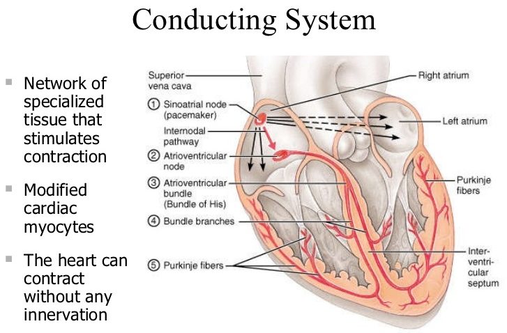
Purkinje fibers provide the heart’s conduction system to perform synchronized contractions of its ventricles, and are, consequently, necessary for keeping a steady heart rhythm. It is committed to a systematic rhythm of the heart. They extend from the interventricular septum, through the papillary muscles and laterally up the walls of the ventricles. A Czech anatomist and physiologist, who discovered the fibers in 1839.
Purkinje Fibers Histology
Purkinje fibers are networks of fibers that are a unique cardiac end-organ. Histologic examination exhibits that these fibers are split in ventricles walls. The electrical origin of atrial Purkinje fibers enters from the sinoatrial(SA) node. The Purkinje fibers are precisely shielded from each other by collagen or the cardiac skeleton.
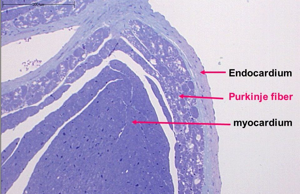
The Purkinje fibers are different from the surrounding muscle cells because of comparatively fewer myofibrils than other cardiac cells and the carriage of glycogen throughout the nucleus, causes Purkinje fibers to appear, on a slide, lighter and bigger than their neighbors, arrayed along the longitudinal orientation (parallel to the cardiac vector). They are usually binucleated cells.
Purkinje Fibers Function
Purkinje fibers are an indispensable part of the functioning of the heart and are vital for our survival. They play a role of the relaying system of electrical signals in the heart, which manages the rate while the cardiac muscles contract and relax, or the rate at which the heart ‘beats’. Definitely, Purkinje fibers cause the ventricles, the lower two chambers of the heart, to create synchronized contractions of its ventricles.
The Purkinje fibers allow for a coordinated and controlled rhythm of blood circulation. Purkinje fibers comprise a distinct type of cardiac muscles that secure them the numerous efficient and quickest conductors of impulses in the cardiac system. The histology of Purkinje fibers reveals that they have thinner muscle tissues with fewer muscle strands than standard cardiac cells, which gives them appear lighter than different cells in a stained slide.