Muscles of the gluteal region: The gluteal region is located posteriorly to the pelvic girdle, at the proximal end of the femur. It can be described as an area of buttocks. There are two gluteal regions, left and right. The muscles in the gluteal region move the lower limb at the hip-joint.
The muscles of the gluteal region broadly classified into two groups:
Superficial abductors and extenders – Muscles that abduct and extend the femur. Superficial group muscles are- gluteus maximus, gluteus medius, gluteus minimus and tensor fascia lata.
Deep lateral rotators-Muscles that largely act to laterally rotate the femur. Deep lateral rotators muscles are- piriformis, gemellus superior, gemellus inferior, quadratus femoris and obturator internus.
Superficial Muscles
Gluteus Maximus: The gluteus maximus is the main extensor muscle of the hip. The gluteus maximus is the largest and most superficial gluteal region muscles. Its thick fleshy mass, in a quadrilateral shape, makes the distinction of the buttocks.
Origin:
- Outer slope of the dorsal segment of the iliac crest
- Gluteal surface of ilium
- Dorsal surface of lower part of the sacrum
- Side of coccyx
- Sacrotuberous ligament
Insertion:
- Gluteal tuberosity
- Iliotibial tract
Nerve supply: Inferior gluteal nerve
Action:
- Chief extensor of the thigh
- Essential for maintaining an erect posture
- Lateral rotation of the thigh
- Abduction of the thigh
Gluteus Medius: The gluteus medius is a broad, thick, radiating muscle, situated on the outer surface of the pelvis. The gluteus medius posterior third is covered by the gluteus maximus, its anterior two-thirds by the gluteal aponeurosis.
Origin: Gluteal surface of ilium between the anterior and posterior gluteal line
Insertion: Lateral surface of greater trochanter of the femur
Nerve supply: Superior gluteal nerve
Action: Powerful abductors of the thigh
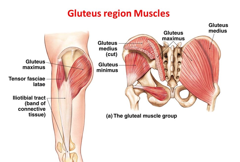
Gluteus Minimus: The gluteus minimus is the tiniest of the three gluteal muscles, the gluteus minimus muscle is situated immediately beneath the gluteus medius.
Origin: Gluteal surface of ilium between the anterior and inferior gluteal line
Insertion: Anterior surface of the greater trochanter of the femur
Nerve supply: Superior gluteal nerve
Action: Powerful abductors of the thigh
Tensor Fasciae Latae: The tensor fasciae latae is a tiny muscle, It is associated with the gluteus maximus in function and structure and is continuous with the iliotibial tract, which connects to the tibia. This band, also described the IT band— located in the thigh and knee. The muscle assists in preserving the balance of the pelvis while walking, standing, or running.
Origin: Anterior surface of the outer lip of iliac crest up to the tubercle
Insertion: Iliotibial tract 3-5cm below the level of the greater trochanter
Nerve supply: Superior gluteal nerve
Action:
- Abductor of thigh
- Medial rotator of thigh
- Extensor of knee joint
Deep Muscles
Piriformis: The piriformis is a muscle in the gluteal region. The piriformis muscle is one of six deep lateral rotators of the hip including is one of only three muscles that attach the legs to the spine.
Origin:
- Pelvic surface of the sacrum
- Upper margin of the greater sciatic notch
Insertion: Apex of the greater trochanter of the femur
Nerve supply: Ventral rami of S1, S2
Action: Lateral rotation of the thigh
Gemellus Superior: The Gemellus superior, the smaller of the two gemellus muscles.The gemellus superior and gemellus inferior help to laterally rotate the extended thigh.
Origin: Upper part of the lesser sciatic notch
Insertion: Medial surface of greater trochanter of the femur
Nerve supply: Nerve to obturator internus
Action: Lateral rotator of thigh at hip joint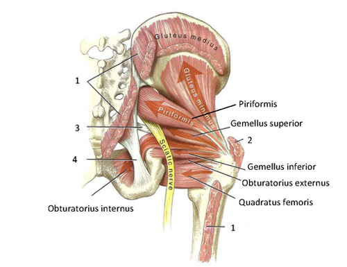
Gemellus Inferior: The Gemellus inferior originates from the upper part of the tuberosity of the ischium, The gemellus superior and the gemellus inferior aid to laterally rotate the extended thigh.
Origin: Lower part of the lesser sciatic notch
Insertion: Medial surface of greater trochanter of the femur
Nerve supply: Nerve to obturator internus
Action: Lateral rotator of thigh at hip joint
Obturator Internus: The obturator internus muscle arises on the medial surface of the obturator membrane, the ischium near the membrane, and the rim of the pubis.
Origin: Pelvic surface of obturator membrane
Insertion: Medial surface of the greater trochanter of the femur
Nerve supply: Nerve to obturator internus
Action: Lateral rotator of thigh at hip joint
Obturator Externus: The obturator externus muscle is a flat, triangular muscle, which covers the outer surface of the anterior wall of the pelvis.
Origin: Outer surface of obturator membrane
Insertion: Medial surface of the greater trochanter
Nerve supply: Posterior division of obturator nerve
Action: Lateral rotator of thigh at hip joint
Quadratus Femoris: The quadratus femoris is a plane, quadrilateral muscle. Positioned on the back side of the hip joint, it is a powerful external rotator and adductor of the thigh but additionally acts to stabilize the femoral head in the Acetabulum.
Origin: Upper part of the outer border of the ischial tuberosity
Insertion: Quadrate tubercle
Nerve supply: Nerve to quadratus femoris
Action: Lateral rotator of thigh at hip joint
Hip Movements
Hip Flexion: Forward and upward movement from the femur through the hip happens in the sagittal plane about a medial-lateral axis.
Hip Flexor Muscles-iliopsoas, rectus femoris, sartorius, pectineus.
Hip Extension: Upward movement toward the rear of the body of the femur at the hip occurring in the sagittal plane.
Hip Extensor Muscles-gluteus Maximus; semimembranosus, semitendinosus and biceps femoris (the hamstrings).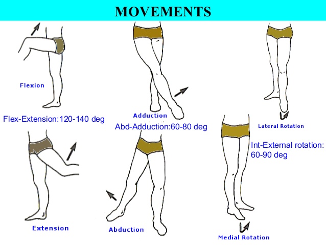
Hip Abduction: Movement of the femur on the hip in a direction away from the midline of the body in the frontal plane.
Hip Abductor Muscles-gluteus medius, gluteus minimus and tensor fascia latae.
Hip Adduction: Movement of the femur on the hip in a direction toward the midline of the body in the frontal plane.
Hip Adductor Muscles-adductors longus, brevis and magnus, pectineus, and gracilis.
Hip Internal Rotation: Rotation of the femur toward the midline of the body in the transverse plane.
Hip Internal Rotator Muscles-anterior fibers of gluteus medius and minimus, tensor fascia latae.
Hip External Rotation: Rotation of the femur away from the midline of the body in the transverse plane.
Hip External Rotator Muscles-biceps femoris, gluteus maximus, helped by the obturators, gemilli and quadratus femoris.