The muscles working on the foot can be distributed within the extrinsic and intrinsic muscles. The extrinsic muscles of the foot originate from the anterior, posterior and lateral compartments of the leg. The extrinsic muscles are largely responsible for eversion, inversion, dorsiflexion, and plantarflexion of the foot.The intrinsic muscles of the foot are placed within the foot and are liable for the fine motor actions of the foot, for instance, movement of individual digits.
Dorsal Aspect
The extrinsic muscles connect to the dorsum of the foot, are only two intrinsic muscles – the extensor hallucis brevis and the extensor digitorum brevis.
Extensor Hallucis Brevis: The extensor hallucis brevis of the foot that assists to extend the big toe. The extensor hallucis brevis is lateral to extensor hallucis longus and medial to the extensor digitorum longus.
- Origin : The extensor hallucis brevis originates from the calcaneus, the interosseous talocalcaneal ligament, and the inferior extensor retinaculum.
- Insertion : The muscle inserts to the base of the proximal phalanx of the great toe.
- Function : The extensor hallucis brevis assists the extensor hallucis longus in extending the great toe at the metatarsophalangeal joint.
- Nerve Supply : Deep fibular nerve.
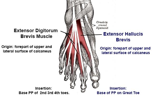
Extensor Digitorum Brevis: The extensor digitorum brevis muscle of the foot that helps extend digits 1 through 4.The extensor digitorum brevis muscle occupies deep to the tendon of the extensor digitorum longus.
- Origin: The extensor digitorum brevis originates from the calcaneus, the interosseous talocalcaneal ligament, furthermore the inferior extensor retinaculum.
- Insertion: It inserts to the to the long extensor tendons of the four lateral digits.
- Function: The extensor digitorum brevis assists the extensor digitorum longus in extending the lateral four toes at the metatarsophalangeal and interphalangeal joints.
- Nerve Supply: Deep fibular nerve.
Plantar Aspect of the Foot
10 intrinsic muscles are found in the sole of the foot. The muscles act collectively to support the arches of the foot, and separately to control the movement of the digits. All the foot muscles are nerve supplied either by the lateral plantar nerve or medial plantar nerve, both are branches of the tibial nerve.
The foot muscles of the plantar aspect are in four layers(superficial to deep).
First Layer of the Plantar Aspect of the Foot
- Abductor Hallucis
- Flexor Digitorum Brevis
- Abductor Digiti Minimi
Abductor Hallucis: The abductor hallucis is positioned on the medial side of the sole.
- Origin: The abductor hallucis arises from the medial tubercle of the calcaneus, the flexor retinaculum, and the plantar aponeurosis.
- Insertion: It inserts to the medial base of the proximal phalanx of the great toe.
- Function: Abducts and flexes the great toe.
- Nerve Supply: Medial plantar nerve.
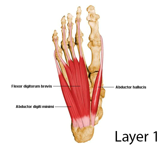
Flexor Digitorum Brevis: The flexor digitorum brevis is positioned laterally to the abductor hallucis. It rests in the middle of the sole, sandwiched within the plantar aponeurosis and tendons of flexor digitorum longus.
- Origin: The flexor digitorum brevis originates from the medial tubercle of the calcaneus and the plantar aponeurosis.
- Insertion: It inserts to the middle phalanges of the lateral four digits.
- Function: Flexes the lateral four digits at the proximal interphalangeal joints.
- Nerve Supply: Medial plantar nerve.
Abductor Digiti Minimi: The abductor digiti minimi is positioned on the lateral side of the foot.
- Origin: The abductor digiti minimi originates from the medial and lateral tubercles of the calcaneus and the plantar aponeurosis.
- Insertion: It inserts to the lateral base of the proximal phalanx of the 5th digit.
- Function: Abducts and flexes the 5th digit.
- Nerve Supply: Lateral plantar nerve.
Second Layer of the Plantar Aspect of the Foot
- Quadratus Plantae
- Lumbricals
Quadratus Plantae: The quadratus plantae is positioned superior to the flexor digitorum longus tendons. The quadratus plantae is parted from the first layer of the foot muscles by the lateral plantar vessels and nerve.
- Origin: The quadratus plantae originates from the medial and lateral plantar surface of the calcaneus.
- Insertion: It inserts to the tendons of flexor digitorum longus.
- Function: Assists flexor digitorum longus in flexing the lateral four digits.
- Nerve Supply: Lateral plantar nerve.
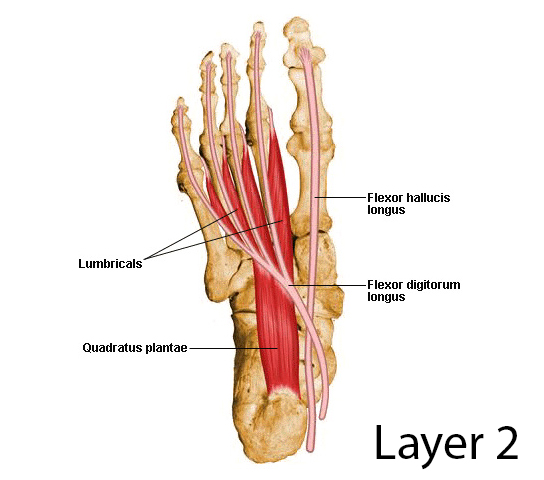
Lumbricals: Lumbrical muscles are of four-foot muscles in the foot. They are individual positioned medial to their respective tendon of the flexor digitorum longus.
- Origin: Lumbrical muscles originate from the tendons of flexor digitorum longus.
- Insertion: It inserts to the extensor hoods of the lateral four digits.
- Function: Flexes at the metatarsophalangeal joints, while extending the interphalangeal joints.
- Nerve Supply: The most medial lumbrical is supplied by the medial plantar nerve. The remaining three are by the lateral plantar nerve.
Third Layer of the Plantar Aspect of the Foot
- Flexor Hallucis Brevis
- Adductor Hallucis
- Flexor Digiti Minimi Brevis
Flexor Hallucis Brevis: The flexor hallucis brevis is positioned on the medial side of the foot.
- Origin: The flexor hallucis brevis originates from the plantar surfaces of the cuboid and lateral cuneiforms, and from the tendon of the posterior tibialis tendon.
- Insertion: It inserts to the base of the proximal phalanx of the great toe.
- Function: Flexes the proximal phalanx of the great toe at the metatarsophalangeal joint.
- Nerve Supply: Medial plantar nerve.
Adductor Hallucis: The adductor hallucis is positioned laterally to the flexor hallucis brevis. The adductor hallucis consists of an oblique and transverse head.
- Origin: The oblique head of the adductor hallucis originates from the bases of the 2nd, 3rd, and 4th metatarsals. The transverse head of the adductor hallucis originates from the plantar ligaments of the metatarsophalangeal joints.
- Insertion: Both heads of the adductor hallucis insert to the lateral base of the proximal phalanx of the great toe.
- Function: Adduct the great toe. Assists in forming the transverse arch of the foot.
- Nerve Supply: Deep branch of the lateral plantar nerve.
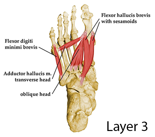
Flexor Digiti Minimi Brevis: The flexor digiti minimi brevis is positioned on the lateral side of the foot, below the metatarsal of the little toe.
- Origin: The flexor digiti minimi brevis originates from the base of the fifth metatarsal.
- Insertion: It inserts to the base of the proximal phalanx of the fifth digit.
- Function: Flexes the proximal phalanx of the fifth digit.
- Nerve Supply: Superficial branch of lateral plantar nerve.
Fourth Layer of the Plantar Aspect of the Foot
The plantar and dorsal interossei cover the fourth plantar muscle layer. The plantar interossei muscle have a unipennate morphology, while the dorsal interossei muscles are bipennate.
- Plantar Interossei
- Dorsal Interossei
Plantar Interossei: There are three plantar interossei muscles, which are positioned among the metatarsals. Individually originate from a single metatarsal.
- Origin: Plantar Interossei originates from the medial side of metatarsals three to five.
- Insertion: It inserts to the medial sides of the phalanges of digits three to five.
- Function: Adduct digits three to five and flexes the metatarsophalangeal joints.
- Nerve Supply: Lateral plantar nerve.
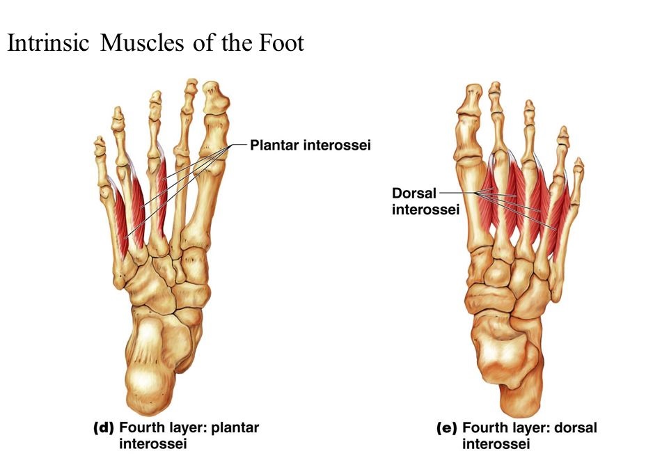
Dorsal Interossei: There are four dorsal interossei muscles, which are positioned within the metatarsals. All arises from two metatarsals.
- Origin: Dorsal Interossei originates from the sides of metatarsals one to five.
- Insertion: The first Dorsal Interossei muscle inserts to the medial side of the proximal phalanx of the second digit. The second Dorsal Interossei muscle to fourth interossei inserts to the lateral sides of the proximal phalanxes of digits two to four.
- Function: Abduct digits two to four and flex the metatarsophalangeal joints.
- Nerve Supply: Lateral plantar nerve.
Movements of the Foot & Ankle
Inversion (Supination):
- The inversion and eversion movements of the foot proceeding the talus.
- The inversion movement is the medial border of the foot where the sole faces medially.
- Muscles acting on Inversion(ROM = 30°)-Tibialis anterior, Tibialis posterior, Flexor hallucis longus & Flexor digitorum longus.
Eversion (Pronation):
- The eversion movement is the lateral border of the foot where the sole faces laterally.
- Muscles acting on Eversion(ROM = 20°)- Peroneus longus,Peroneus brevis & Peroneus tertius.
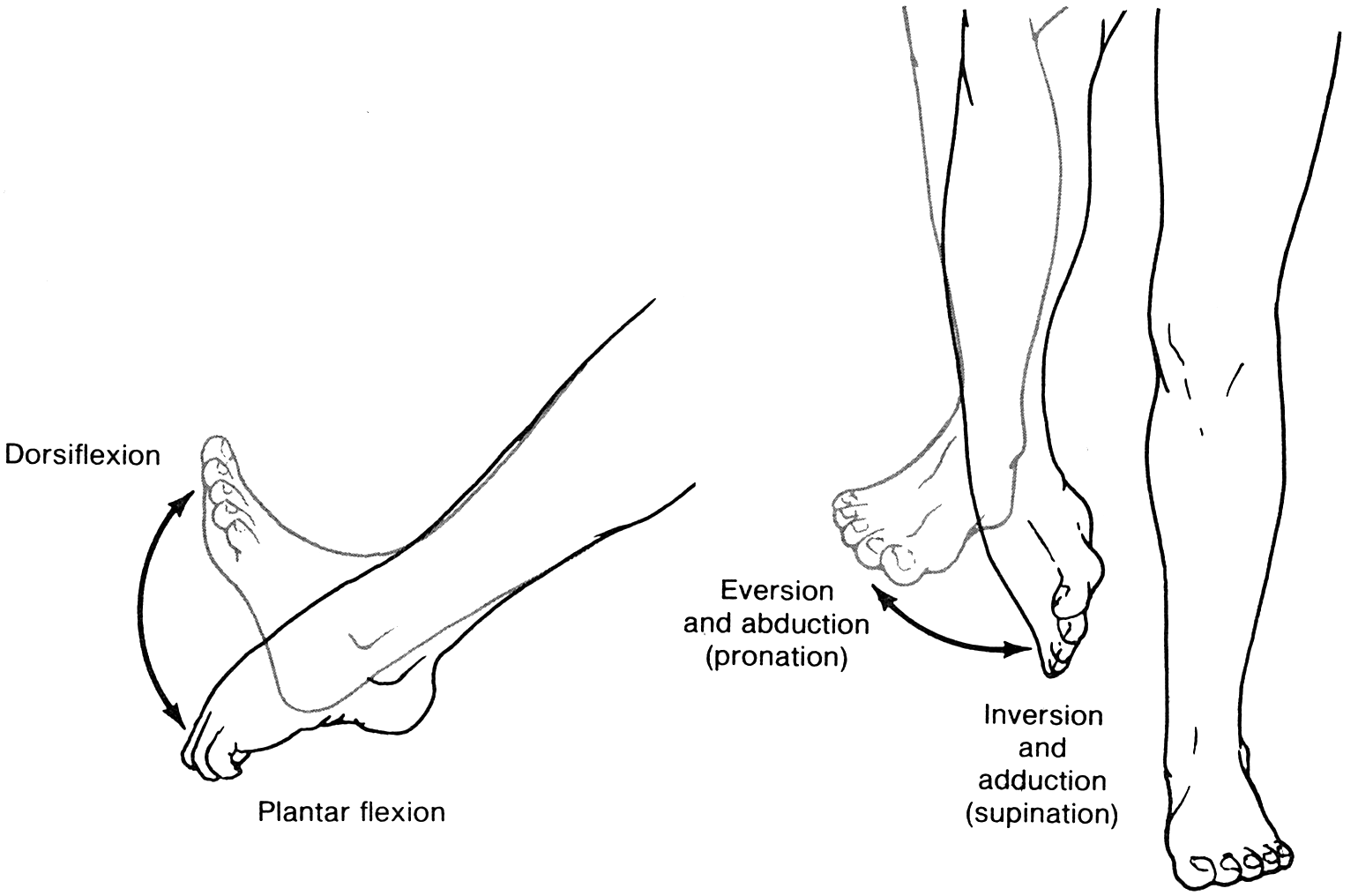
Dorsiflexion:
Dorsiflexion of foot involves moving the top of the foot towards the shin.
The dorsiflexors muscles combine-
- Tibialis anterior
- Extensor Hallucis Longus
- Extensor Digitorum Longus
- Peroneus Tertius
Plantar Flexion:
Plantar flexion of the foot is the opposite movement of the Dorsiflexion otherwise known as pointing your toes down.
The Plantar flexor muscles combine-
- Gastrocnemius
- Soleus
- Plantaris
- Flexor hallucis longus
- Flexor digitorum longus
- Tibialis posterior
- Peroneus longus
- Peroneus brevis
The toes can be flexed, extended, abducted and adducted. Movement of the midtarsal joint is the rotation.
Common Foot Problems That Cause Pain:
Extensor Tendonitis, Tibial Tendonitis, Ball of Foot Pain, Arch of Foot Pain, Plantar Fasciitis, Bunion, Heel Spurs & Diabetic Neuropathy.