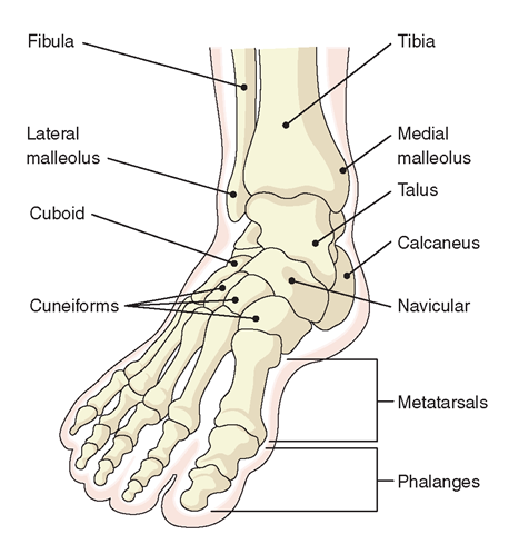
Foot Bones: There are thirty-three-foot bones making the foot inhuman. They are designed to be strong, durable and yet flexible adequately for movement. The foot bones are often grouped into 3 sections:
- Hindfoot
- Midfoot
- Forefoot
Hindfoot of the Foot bones
The hindfoot forms the heel and ankle. There are two joints in the hindfoot, the ankle joint, and the subtalar joint. The talus bone supports the leg bones (tibia and fibula), forming the ankle. The calcaneus (heel bone) is the largest bone in the foot. The ankle joint allows the up and down motion of the ankle, the subtalar joint allows the side to side movement.
1) Tibia
2) Fibula
3) Talus
4) Calcaneus
Tibia
The tibia is the foremost bone of the leg. The tibia has a proximal and distal end and a shaft, connecting at the knee in proximal furthermore ankle joints in the distal end. The tibia is the second largest bone in the body.
The ankle joint contains the tibia, fibula and the talus bones. The tibia forms the top and inner side of the ankle joint whether it meets the talus bone of the foot. Towards the ground of the tibia, the bone increases outwards making the medial malleolus, the notable part on the inner side of the ankle.
Fibula
The fibula is the long, thin and lateral bone of the leg that runs down the outer side of the lower leg. The fibula plays a notable role in stabilizing the ankle joint and supporting the muscles of the lower leg. At the bottom it extends outwards, forming a bony prominence known as lateral malleolus. It extends slightly lower than the medial malleolus forming the outer side of the ankle joint where it connects with the talus.
Talus
The talus forms the lower part of the ankle joint, joining with the tibia and fibula and the top of the subtalar joint, connecting with the calcaneus. The talus sits above the heel bone (calcaneus). No muscles attach to the talus. The talus held in place by the bones enclosing it and various ligaments.
The talus and calcaneus make the subtalar joint, that is important for walking, especially on rough ground.
Calcaneus
The calcaneus is known as the heel bone. It is the largest foot bones and quadrangular shape. The calcaneus is commonly fractured usually from a high fall. The Achilles tendon connects calcaneus linking the calf muscles to back of the heel. Collectively, the calcaneus and the talus make the subtalar joint. The subtalar joint provides side-to-side movement of the hindfoot and is particularly necessary for balance on rough surfaces.
Midfoot of the Foot bones
The midfoot is a pyramid-like arrangement of bones that sit unitedly to form the foremost shape of the foot and the foot arches. These include
1) Navicular
2) Cuboid
3) Cuneiform Bones
Navicular
The navicular is found on the inner side of the foot. It articulates proximally with the talus, distally with the three cuneiform bones, and laterally with the cuboid. The tibialis posterior attaches to the navicular bone.
Cuboid
The cuboid bone in the foot is found on the outer side of the foot in the distal row of the tarsus. At the back, cuboid bone connects to the heel bone, identified as the calcaneocuboid joint. On the medial side, cuboid bone articulates with the navicular and third cuneiform on the medial side. At the front, cuboid bone makes the lateral link within the tarsal and metatarsal bones, identified as the tarsometatarsal joints.
Cuneiform Bones
There are three cuneiform bones:
- Medial Cuneiform: on the inner
- Intermediate Cuneiform: in the middle
- Lateral Cuneiform: on the outer side
The medial cuneiform is the largest of the cuneiforms. It is located at the medial side of the foot, anterior to the navicular bone, posterior to the first metatarsal and lateral to the intermediate cuneiform. The medial cuneiform articulates with four bones: the navicular, second cuneiform, and first and second metatarsals. The fibularis longus and tibialis anterior muscle inserts at the medial cuneiform bone.
The intermediate cuneiform is like a wedge-shaped, with the thin end pointing downwards. The intermediate cuneiform is located within the other two cuneiform bones and articulates with the navicular posteriorly and the second metatarsal anteriorly
The lateral cuneiform is also wedge-shaped, the base being uppermost. The lateral cuneiform keeps the center of the front row of the tarsal bones, within the intermediate cuneiform medially, the cuboid laterally, the navicular posteriorly and the third metatarsal in front. The tibialis posterior muscle inserts at the medial cuneiform, while the flexor hallucis brevis muscle originates from it.
Forefoot of the Foot Bones
The forefoot contains the five long metatarsal bones and the fourteen small toe bones known as the phalanges.
1) Metatarsal Bones
2) Phalanges
The most prevalent difficulties concerning the foot and ankle bones are fractures, abnormal positioning ( flat feet) and unnecessary bone growth (bunions and bone spurs).
Metatarsal Bones
The metatarsals are positioned between the tarsals and phalanges. They are calculated I-V (medial to lateral). The metatarsals are the long bones that
Proximally: Tarsometatarsal joint – within the metatarsal bases and the bones of the cuneiforms or cuboid.
Distally: Metatarsophalangeal joint – within the metatarsal head and the bones of the proximal phalanx.
The first metatarsal bone, the shortest and strongest metatarsal, connects to the big toe. The second metatarsal is the largest. These foot bones get shorter towards the fifth metatarsal bone which connects to the little toe.
Each metatarsal has a head, body, and base. They articulate proximally to the tarsal bones. The first metatarsal connects to the medial and intermediate cuneiform, the second metatarsal bone to all three cuneiforms, the third metatarsal bone to the lateral cuneiform, the fourth metatarsal bone to the lateral cuneiform and cuboid bones and the fifth metatarsal to the cuboid bone.
Fractures of the metatarsal bones are often seen in football players.
Phalanges
The phalanges hold the little foot bones that make up the toes. The big toe is formed up of two phalanges, whilst the other four toes made up of three phalanges:
1) Proximal Phalanges that connect with the metatarsals, known as the metatarsophalangeal joints (MTP). The Proximal Phalanges tend to be the largest of all the phalanges.
2) Middle Phalanges are the small phalanges in the middle of the toes. The big toe has not a middle phalanx.
3) Distal Phalanges are the toe bones furthest away from the body.
The joints connecting the proximal and middle phalanges are identified as the proximal interphalangeal joints (PIP) and the joints connecting the middle and distal phalanges are identified as the distal interphalangeal joints (DIP).
Foot Bones Common Problems
The common Foot Bones problems affecting the foot bones are the injury which can change the position of the joints:
Bunions
Commonly attack the big toe, the symptom which is the toe deviating inwards producing a lump on the outer side of the big toe.
Fractures
If too much force is placed within the foot bones, either throughout a one-off injury or as a consequence of repetitive stress, the bones can fracture.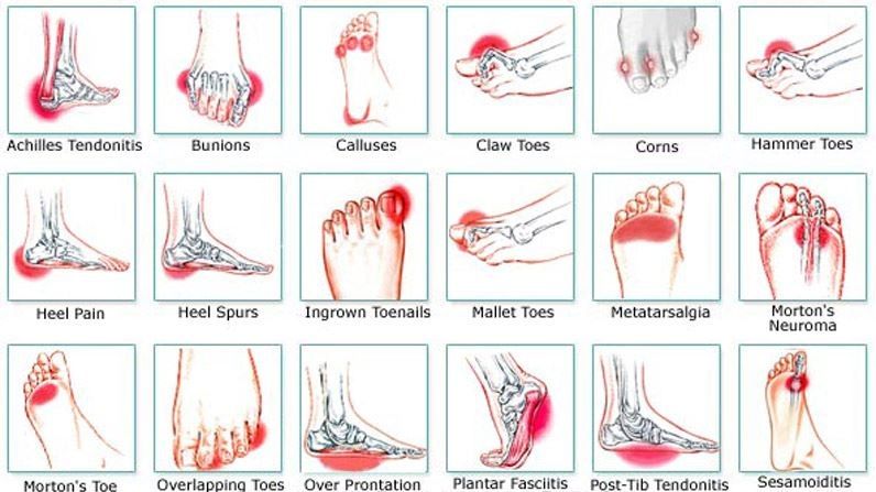
Bone Spurs
These develop repeated friction on the bone.
Mallet, Hammer & Claw Toe
These problems affecting the toe joints. The toe joints get forced into the wrong position in various forms causing the toes to bend.
Turf Toe
This problem in athletes affecting the big toe. It creates pain, inflammation, and stiffness.