Eye Muscles: There are seven extraocular eye muscles that are present in the eye socket that join the eye to move it. These muscles control to move the eye from side to side, up, down and rotate the eye.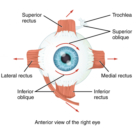
Extraocular Muscles
The extraocular muscles are placed in the orbit but are extrinsic and separate from the eyeball itself. They work to control the movements of the eyeball including the superior eyelid. There are seven extraocular muscles-
Levator palpebrae superioris: Responsible for superior eyelid movement,
Recti and oblique muscles: Responsible for eye movement.
Voluntary Muscles:
- levator Palpebrae Superior
Four Recti Muscles:
- Superior rectus
- Inferior rectus
- Medial rectus
- Lateral rectus
Two Obliques Muscles:
- Superior oblique
- Inferior oblique
levator Palpebrae Superior: The levator palpebrae superioris is the only muscle which is involved in raising the superior eyelid. A small portion of this muscle contains smooth muscle fibers – known as the superior tarsal muscle. The superior tarsal muscle is innervated by the sympathetic nervous system.
Origin: Originates from the lesser wing of the sphenoid bone, immediately above the optic foramen.
Insertion: The levator palpebrae superioris is inserted to the superior tarsal plate of the upper eyelid.
Function: Elevates the upper eyelid.
Nerve Supply: The levator palpebrae superioris is supplied by the oculomotor nerve (CN III). The superior tarsal muscle is by the sympathetic nervous system.
Six skeletal muscles surround and produce various eye movements. Four of the extraocular muscles originated from a tendinous band surrounding the optic nerve. This band, known as the annulus of Zinn.
Recti Muscles:
The superior rectus is a thin muscle and forms a straight muscular band between the eye and the annulus of Zinn. The inferior, medial, and lateral rectus muscles are almost identical to the superior rectus muscle, except they insert on the inferior, medial, and lateral edges of the eye.
Superior Rectus:
Origin: Originates from the superior part of the common tendinous ring.
Insertion: Superior Rectus inserts to the superior and anterior aspect of the sclera.
Function: the Main movement is elevation. Also adduction and medial rotation of the eyeball.
Nerve Supply: Oculomotor nerve (CN III).
Inferior Rectus:
Origin: Originates from the inferior part of the common tendinous ring.
Insertion: Inferior Rectus inserts to the inferior and anterior aspect of the sclera.
Function: the Main movement is depression, Also adduction and lateral rotation of the eyeball.
Nerve Supply: Oculomotor nerve (CN III).
Medial Rectus:
Origin: Originates from the medial part of the common tendinous ring,
Insertion: Medial Rectus inserts to the anterior-medial aspect of the sclera.
Function: Adducts the eyeball.
Nerve Supply: Oculomotor nerve (CN III).
Lateral Rectus:
Origin: Originates from the lateral part of the common tendinous ring.
Insertion: Lateral Rectus inserts to the anterior-lateral aspect of the sclera.
Function: Abducts the eyeball.
Nerve Supply: Abducens nerve (CN VI).
Oblique Muscles:
The superior oblique muscle originates at the back of the orbit, called the trochlea, on the upper, nasal wall of the orbit. The muscle inserts on the lateral, posterior part of the globe. The superior oblique muscle pulls the eye downward and laterally.
The inferior oblique, which originates at the lower front of the nasal orbital wall, and inserts on the lateral, posterior part of the globe. The inferior oblique pulls the eye upward and laterally.
Superior Oblique:
Origin: Originates from the body of the sphenoid bone.
Insertion: Superior Oblique attaches to the sclera of the eye, posterior to the superior rectus.
Function: Depresses, abducts and medially rotates the eyeball.
Nerve Supply: Trochlear nerve (CN IV).
Inferior Oblique:
Origin: Originates from the anterior aspect of the orbital floor.
Insertion: Inferior Oblique inserts to the sclera of the eye, posterior to the lateral rectus.
Function: Elevates, abducts and laterally rotates the eyeball.
Nerve Supply: Oculomotor nerve (CN III).
Involuntary Muscles:
- Superior tarsal muscles
- Inferior tarsal muscles
Blood Supply
The extraocular muscles are supplied mainly by branches of the ophthalmic artery. Supplementary branches of the ophthalmic artery combine the ciliary arteries, which branch into the anterior ciliary arteries. Each rectus muscle takes blood from two anterior ciliary arteries, except for the lateral rectus muscle, which takes blood from only one. Branches of the infraorbital artery supply the inferior rectus and inferior oblique muscles.