Back Muscles: The muscles of the back that work together to support the spine, help keep the body upright and allow twist and bend in many directions. The back muscles can be three types.
a. Superficial Back Muscles
b. Intermediate Back Muscles and
c. Deep Back Muscles
Superficial Back Muscles
- Action
- Movements of the shoulder.
Intermediate Back Muscles
- Action
- Movements of the thoracic cage.
Deep Back Muscles
- Action
- Movement of the vertebral column
The deep muscles develop in the back called intrinsic muscles.
The superficial and intermediate muscles do not develop in the back so-called extrinsic muscles.
Superficial Back Muscles
These muscles are situated underneath the skin and superficial fascia. They arise from the vertebral column and inserted into the bones of the shoulder such as
- Clavicle
- Scapula and
- Humerus
All superficial muscles associated with movements of the upper limb. Superficial muscles are given below-
- Trapezius
- Latissimus dorsi
- Levator Scapulae
- Rhomboids
Trapezius and latissimus dorsi lie in the very much superficially. Trapezius covering levator scapulae and the rhomboids.
Trapezius: The trapezius is a broad, flat and triangular muscle. The muscles on each side form a trapezoid shape. It is the most superficial of all the back muscles.
- Origin:
- Occipital protuberance
- Ligamentum nuchae
- The spine of 7th cervical vertebra
- Spine of all thoracic vertebra
- Insertion:
- Upper fibers into posterior border of the lateral third of the clavicle
- Middle fibers into the medial margin of the acromion and upper lip of the crest of the spine of the scapula
- Lower fibers on the deltoid tubercle
- Nerve supply:
- Spinal part of the accessory nerve is motor
- Branches from C3, C4 are proprioceptive
- Action:
- Upper fibers elevate the scapula
- Middle fibers retract the scapula
- Lower fibers pull the scapula inferiorly.
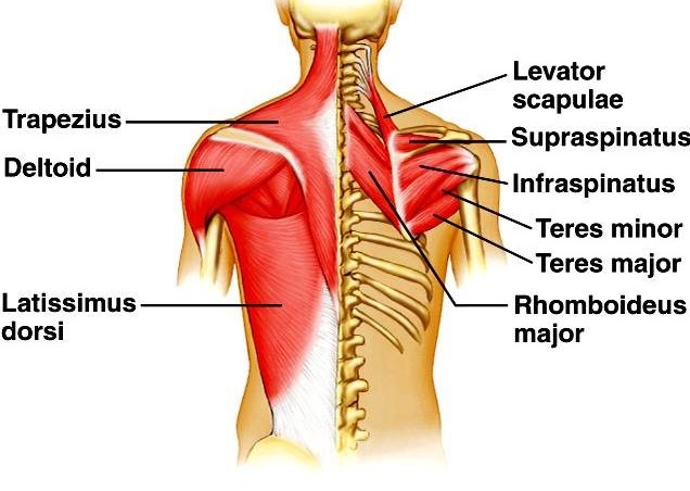
Latissimus dorsi: The latissimus dorsi originates from the lower part of the back, where it covers a wide area.
- Origin:
- Posterior one-third of the outer lip of iliac crest
- Posterior layer of lumbar fascia
- Spines of T7-T12 Vertebra
- Lower four ribs
- Inferior angle of the scapula
- Insertion:
- The bicipital groove of the humerus
- Nerve supply:
- Thoracodorsal nerve ( nerve to latissimus dorsi)
- Action:
- Adduction, extension and medial rotation of the shoulder
Levator scapulae: The levator scapulae is a small strap-like muscle. It begins in the neck and descends to attach to the scapula.
- Origin:
- Transverse process of first four cervical vertebrae (C1 –C4 )
- Insertion:
- Superior angle and upper part of medial border of scapula
- Nerve supply:
- Dorsal scapular nerve
- Action:
- Elevation of the scapula
Rhomboids: There are two muscles.
- Rhomboideus minor
- Rhomboideus major
Rhomboideus minor is situated above major.
Rhomboideus minor:
- Origin:
- The lower part of ligamentum nuchae
- The spine of C7 and T1 vertebra
- Insertion:
- Upper 1/3rd of medial border of scapula
- Nerve supply:
- Dorsal scapular nerve
- Action:
- Retraction rotation of the scapula
Rhomboideus major:
- Origin:
- Spine of the T2-T5 vertebra
- Insertion:
- Medial border of scapula between the spine of scapula and inferior angle.
- Nerve supply:
- Dorsal scapular nerve
- Action:
- Retraction rotation of the scapula
Intermediate Back Muscles
- Serratus posterior superior
- Serratus posterior inferior
Serratus posterior superior: The serratus posterior superior is a thin muscle, situated at the upper and back part of the thorax, deep to the muscles of the rhomboid.
- Origin:
- Spinous processes and supraspinous ligaments of C7-T2
- Insertion:
- Posterior aspect of ribs 2-5
- Function:
- Assists forced inspiration
- Nerve Supply:
- Anterior primary rami (T2-5)
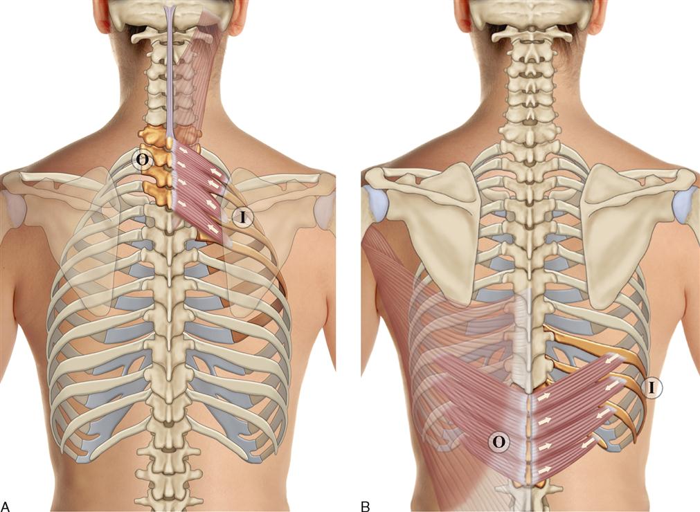
Serratus posterior inferior: The serratus posterior inferior is one of the back’s two intermediate muscles.
- Origin:
- The serratus posterior inferior originates from the spinal processes of vertebrae T11 to T12 and L1 to L2.
- Insertion:
- Inserts into the lower borders of ribs 9-12.
- Function:
- The function of the serratus posterior inferior muscle depresses thoracic ribs 9-12, assisting with forced exhaling.
- Nerve Supply:
- Intercostal nerves.
Deep Back Muscles
The deep back muscles have three layers-
a.Superficial
b.Intermediate
c.Deep.
Superficial Deep Back Muscles:
Action: Movement of the head and neck
Splenius Capitis:
- Origin:
- ligamentam nuchae
- Spinus process of C7 –T3/T4 vertebrae
- Insertion:
- Mastoid process
- The occipital bone of the skull
- Nerve supply:
- Posterior rami of spinal nerves C3 and C4.
- Actions:
- Rotate head to the same side.
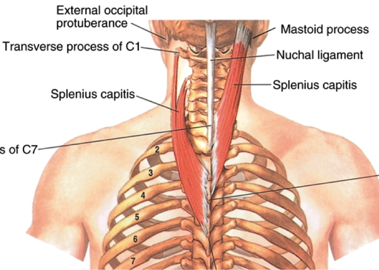
Splenius cervicis:
- Origin:
- Spinus process of T3-T6 vertebrae
- Insertion:
- Transverse processes of C1-C4
- Nerve supply:
- Posterior rami of the lower cervical spinal nerves.
- Action:
- Rotate head to the same side.
Intermediate Deep Back Muscles:
There are in three muscle.
Iliocostalis: The muscle is situated laterally within the erector spinae.The muscle is related to the ribs. The muscle divided into three parts.
- Lumborum
- Thoracis and
- Cervicis
- Origin:
- Common tendinous origin
- Insertion:
- The costal angle of the ribs
- Cervical transverse processes
- Nerve supply:
- Posterior rami of the spinal nerves.
- Action:
- Unilaterally to laterally flex the vertebral column
- Bilaterally to extend the vertebral column and head.
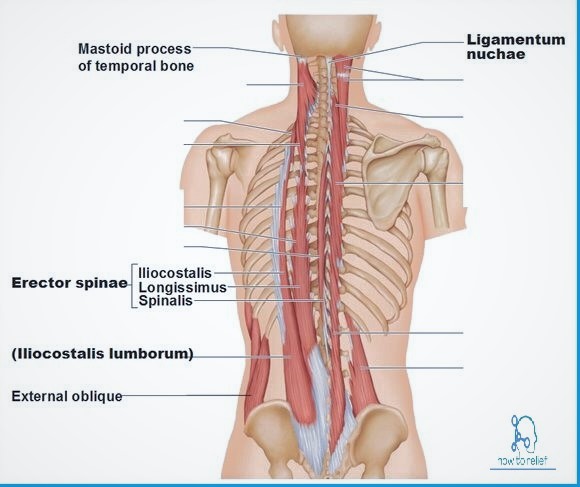
Longissimus: The muscle is located between the iliocostalis and spinalis.This muscle is the largest of the three columns.
The muscle has three parts:
- Thoracic
- Cervicis and
- Capitis.
- Origin:
- Tendinous origin
- Insertion:
- Lower ribs
- Transverse processes of C2-T12
- Mastoid process of the skull
- Nerve supply:
- Posterior rami of the spinal nerves.
- Action:
- Unilaterally to laterally flex the vertebral column.
- Bilaterally to extend the vertebral column and head.
Spinalis: The muscle is situated medially within the erector spinae.The muscle is the smallest of the three muscle columns.
The muscle has three part.
- Thoracic
- Cervicis and
- Capitis
- Origin:
- Common tendinous origin
- Insertion:
- Spinous processes of C2, T1-T8
- The occipital bone of the skull
- Nerve supply:
- Posterior rami of the spinal nerves.
- Action:
- Unilaterally to laterally flex the vertebral column.
- Bilaterally to extend the vertebral column and head.
These three muscles are called erector spinae
Most Deep Back Muscles: These muscles are situated underneath the erector spinae.They are a group of short muscles.
Related to transverse and spinous processes of the vertebral column.
The deep back muscles are:
Semispinalis: The muscle is the most superficial of the deep intrinsic muscles.The muscle has three parts.
- Thoracic
- Cervicis and
- Capitis.
- Origin:
- Transverse processes of C4-T10
- Insertion:
- Spinous processes of C2-T4
- The occipital bone of the skull
- Nerve supply:
- Posterior rami of the spinal nerves.
- Action:
- Extends and contralaterally rotates the head and vertebral column.
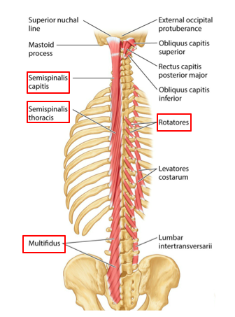
Multifidus: The muscle is situated underneath the semispinalis muscle. This muscle is best developed in the lumbar area.
- Origin:
- Sacrum
- Posterior iliac spine
- Common tendinous origin of the erector spinae
- Mamillary processes of the lumbar vertebra
- Transverse processes of T1-T3 and
- Articular processes of C4-C7.
- Insertion:
- Spinous processes of the vertebrae.
- Nerve supply:
- Posterior rami of the spinal nerves.
- Action:
- Stabilizes the vertebral column.
Rotatores: The muscles are most prominent in the thoracic region.
- Origin:
- Vertebral transverse processes.
- Insertion:
- Lamina and spinous processes of the immediately superior vertebrae.
- Nerve supply:
- Posterior rami of the spinal nerves.
- Action:
- Stabilizes the vertebral column
- Proprioceptive function.
Quadratus Lumborum: The quadratus lumborum is the most posterior deep back muscles of the posterior abdominal wall. The quadratus lumborum is the deepest abdominal muscle and commonly referred to as a back muscle.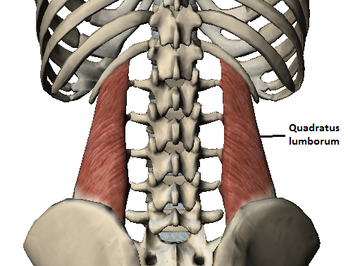
- Origin:
- Posterior Iliac Crest
- Insertion:
- The medial half of 12th rib and transverse processes of lumbar vertebrae.
- Nerve Supply:
- Subcostal nerve (T12), Iliohypogastric and Ilioinguinal nerve (both from L1) & Branches from the ventral rami (L2 and L3)
- Function:
- Quadratus Lumborum fixes the12th rib to stabilize diaphragm attachments during inspiration, Lateral flexes the vertebral column & Extends lumbar vertebrae

