Neck Muscles: Neck muscles are of four types.
- Suboccipital muscles
- Suprahyoid muscles
- Infrahyoid muscles
- Scalene muscles
These four muscles are described below:
Suboccipital Muscles
The suboccipital muscles are a group of four muscles that located under the occipital bone. These muscles are located in the sternocleidomastoid, Trapezius, splenius and semispinalis muscles. They are collectively acting to extend and rotate the head.
Suboccipital muscles are of four types. They are given below:
- Rectus capitis posterior major
- Rectus capitis posterior minor
- Obliquus capitis superior( superior oblique)
- Obliquus capitis inferior( inferior oblique)
Rectus capitis posterior major: The rectus capitis posterior major is located laterally to the rectus capitis posterior minor. It comprises the posterosuperior boarder of the suboccipital triangle.
Origin: Spine of the axis
Insertion: Lateral part of the area below the inferior nuchal line
Nerve supply: Suboccipital nerve
Action:
Mainly postural
Extend the head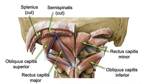
Rectus capitis posterior minor: The rectus capitis posterior minor is the most medial of the suboccipital muscles. There is a connective tissue bridge within this muscle and the dura mater, which may perform a role in cervicogenic headaches.
Origin: Posterior tubercle of the atlas
Insertion: Medial part of the area below the inferior nuchal line
Nerve supply: Suboccipital nerve
Action:
Mainly postural
Extends the head
Obliquus capitis superior: The obliquus capitis superior is located laterally in the suboccipital compartment.
Origin: Transverse process of the atlas
Insertion: the Lateral area between the nuchal line
Nerve supply: Suboccipital nerve
Action:
Mainly postural
Extends the head
Flexes the head laterally
Obliquus capitis inferior: The obliquus capitis inferior is the most inferiorly positioned of the suboccipital muscles.
Origin: Spine of the axis
Insertion: Transverse process of the atlas
Nerve supply: Occipital Nerve
Action:
Mainly postural
Turns chin to the same side
Suprahyoid Muscles
The suprahyoid muscles are a group of four muscles. The suprahyoid muscles located superiorly to the hyoid bone of the neck. They are collectively working to elevate the hyoid bone and involved in swallowing.
The arterial supply to these muscles of the facial artery, occipital artery, and lingual artery.
- Digastric
- Stylohyoid
- Mylohyoid
- Geniohyoid
- Hyoglossus
Digastric (two belly): The digastric muscle is positioned in the neck, beneath the jaw. This muscle assists in opening and closing the jaw.
Origin:
Anterior belly: digastric fossa of mandible
Posterior belly: mastoid notch of temporal bone
Insertion: Both heads meet at the intermediate tendon
Nerve supply:
Anterior belly: nerve to mylohyoid
Posterior belly: facial nerve
Action:
Depresses mandible
Elevates hyoid bone
Stylohyoid: The stylohyoid muscle is a thin muscular strip, that is positioned superiorly to the posterior belly of the digastric muscle.
Origin: Posterior surface of the styloid process
Insertion: Junction of body and greater cornua of the hyoid bone
Nerve supply: Facial nerve
Action:
Pulls hyoid bone upwards and downwards
Flexes the hyoid bone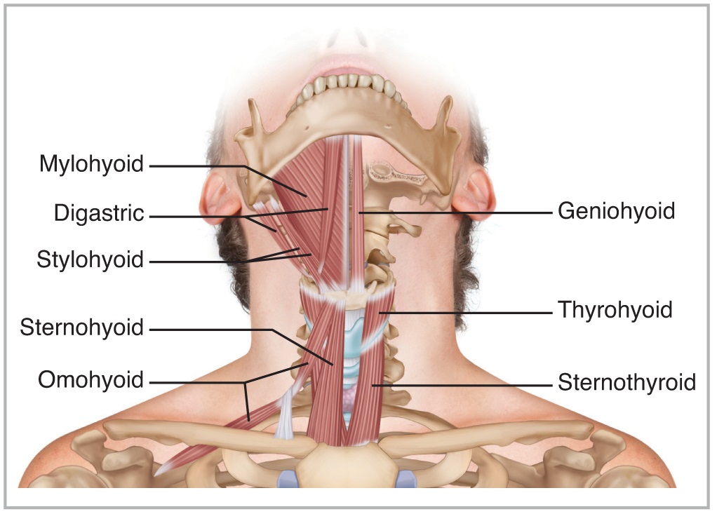
Mylohyoid: The mylohyoid muscle is a paired muscle running from the mandible to the hyoid bone. It forms the floor of the oral cavity and supports the floor of the mouth.
Origin: Mylohyoid line of the mandible
Insertion:
Posterior fibers: the body of hyoid bone
Middle and anterior fibers: between the mandible and hyoid bone
Nerve supply: Nerve to mylohyoid
Action:
Depression of mandible
Elevation of the hyoid bone
Geniohyoid: The geniohyoid muscle is a thin muscle located superior to the medial border of the mylohyoid muscle.
Origin: Inferior mental spine
Insertion: Anterior surface of the body of hyoid bone
Nerve supply: Hypoglossal nerve
Action:
Elevate hyoid bone
Depress mandible
Hyoglossus:
Origin: The Hyoglossus originate from the whole length of greater cornua and lateral part of the body of hyoid bone
Insertion: Side of the tongue
Nerve supply: Hypoglossal nerve
Action:
Depresses tongue
Retracts the protruded tongue
Infrahyoid Muscles
The infrahyoid muscles are a group of four muscles that are positioned inferiorly to the hyoid bone in the neck.
They are classified into two groups.
Superficial muscles: omohyoid and sternohyoid
Deep muscles: sternothyroid and thyrohyoid muscles
Sternohyoid: The sternohyoid muscle is located within the superficial plane.
Origin:
Posterior surface of the manubrium sterni
Adjoining part of the clavicle and the posterior sternoclavicular ligament
Insertion: Medial part of the lower border of hyoid bone
Nerve supply: Ansa cervicalis
Action: Depresses the hyoid bone
Omohyoid: The omohyoid is comprised of two muscle bellies, which are connected by a muscular tendon.
Origin:
Upper border of scapula near the suprascapular notch
Adjoining part of the suprascapular ligament
Insertion: Lower border of hyoid bone lateral to the sternohyoid
Nerve supply:
Superior belly by the superior root of the ansa cervicalis
Inferior belly by ansa cervicalis
Action: Depresses the hyoid bone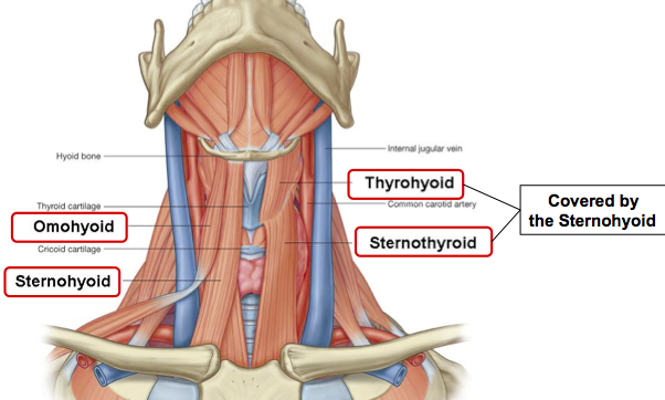
Sternothyroid: The sternothyroid is a muscle in the neck. The sternothyroid muscle is wider and deeper than the sternohyoid.
Origin:
Posterior surface of manubrium sterni
Adjoining part of first costal cartilages
Insertion:
Oblique line on the lamina of the thyroid costal cartilages
Nerve supply: Ansa cervicalis
Action: Depresses the larynx
Thyrohyoid: The thyrohyoid muscle is a short band of muscle, which depresses the hyoid and elevates the larynx.
Origin: Oblique line of thyroid cartilage
Insertion: Lower border of the body and the greater cornua of the hyoid bone
Nerve supply: Hypoglossal nerve
Action:
Depress the thyroid bone
Elevates the larynx
Scalene Muscles
The scalene muscles are three paired muscles. Scalene muscles are located in the lateral aspect of the neck. Collectively Scalene muscles form part of the floor of the posterior triangle of the neck.
The scalene performs as accessory muscles of respiration and produces flexion at the neck.
- Scalenus anterior
- Scalenus medius
- Scalenus posterior
Scalenus anterior: The anterior scalene muscle is one of the lateral muscles of the neck, deep to the prominent sternocleidomastoid muscle.
Origin: Anterior tubercles of transverse processes of C3, C4, C5, and C6.
Insertion: Scalene tubercle
Nerve supply: Ventral rami of C4-C6 nerves
Action:
Anterolateral flexion of the cervical spine
Rotates cervical spine to opposite side
Elevate the first rib
Stabilizes the neck along with other muscles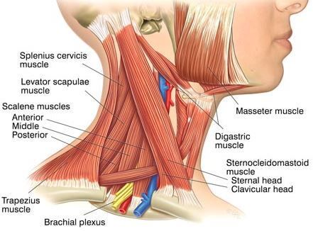
Scalenus medius: The middle scalene is the largest and longest in the Scalene group of the lateral neck muscles.
Origin:
Posterior tubercles of the transverse processes C3-C7 vertebra
Transverse process of the axis and sometimes also of the atlas vertebra
Insertion: the Superior surface of the first rib
Nerve supply: Ventral rami of C3-c8 nerves
Action:
Lateral flexion of the cervical spine
Elevation of the first rib
Scalenus posterior: The posterior scalene is the smallest and deepest of the scalene muscles. It is deeply placed, lying behind Sternocleidomastoid.
Origin: Posterior tubercles of transverse processes of the C4-C6 vertebra
Insertion: Outer surface of the 2nd rib
Nerve supply: Ventral rami of C6-C8 nerves
Action:
Lateral flexion of the cervical spine
Elevation of the 2nd rib
Movements of the Neck
The neck performs essentially six movements. These gently performed movements are:
1)Neck flexion—the neck flexion movement in which the chin is lowered down toward the chest.
Neck flexion muscles-Longus Colli, Longus capitis, Rectus capitis anterior, Sternocleidomastoid.
2)Neck extension—the neck extension is looking upward toward
the ceiling.
Neck extension muscles-Splenius Capitus, Cervicis Muscles, Semispinalis Cervicis, Spinalis Capitus, Semispinalis Capitus, Upper Trapezius, Longissimus Cervicis.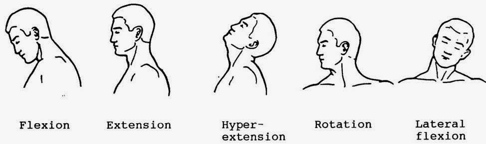
3 and 4)Neck lateral rotation to the left and to the right—the neck lateral rotation are simply direct lateral rotation to either side.
Neck lateral rotation muscles-Sternocleidomastoid, Splenius capitis, Splenius cervicis, Longissimus capitis, Semispinalis thoracis, Semispinalis cervicis, Semispinalis capitis, Rectus capitis posterior major.
5 and 6)Neck lateral flexion—the neck lateral flexion is shoulder through a sideways movement of the neck, directing the ear toward the shoulder tip on both sides.
Neck lateral flexion muscles-Anterior scalene, Middle scalene, Splenius cervicis, Splenius capitis, Rectus capitis lateralis, Longus colli, Intertransversarii.